The mission of Dobrobut is regaining the credibility of Ukrainian medicine. Credibility of the medicine starts from an educated doctor. Therefore, we make sure that our doctors have the most up-to-date knowledge, they are aware of the latest research and possess the best methods of diagnosis, treatment and prevention. We have created Dobrobut Academy to improve the level of knowledge and qualifications of doctors.
Dobrobut Academy is a unique complex educational hub for Ukraine, where doctors, medical personnel and ordinary citizens can gain new medical knowledge and experience. We provide enormous educational opportunities, from obtaining modern medical knowledge to implementing new medical and scientific projects based on Dobrobut medical network.
Go to the Dobrobut Academy website to learn more about all the projects.
Dobrobut Academy has three main fields:
- internship for graduates of medical universities;
- education and training center for those who want to gain quality theoretical knowledge on various types of provision of emergency aid in life-threatening conditions;
- constant professional development for medical staff.
Internship
Dobrobut Academy makes sure that Ukrainian young doctors receive high-quality and modern theoretical, clinical and practical knowledge and confidently start their medical careers. Our internship provides conceptually new approaches to professional education. Interns become full-fledged participants of our large Dobrobut team from the very beginning. They gain practical experience studying at Dobrobut medical centers under the best specialists, the leaders of medical areas.
We want Ukrainian doctors to have the world-class classification.
The internship provides the training in 18 specialties. Dobrobut interns learn to work with medical information systems (MIS), build communication with patients, keep records, provide emergency assistance. Modern equipment, tools, comfortable working environment and personal protective equipment are at their disposal.
Our training programs for interns are developed on the basis of European Union of Medical Specialists (UEMS) programs. Therefore, Dobrobut Academy interns will be able to acquire:
- skills of communication with patients of various categories;
- specifics of working with patients with psychosomatic disorders;
- topical issues of infection control and vaccination;
- legal aspects of the doctor’s work;
- toxicology.
We provide versatile multidisciplinary training. Our interns have the opportunity to work not only in their chosen specialty, but also to adopt the experience of specialists in related medical fields, gaining more knowledge and broadening their “medical horizons”.
Internship fields of Dobrobut Academy
The interns of all medical fields gain practical and theoretical knowledge in the following specialties:
- Anesthesiology and intensive care
Interns in this specialty will, among other things, be able to: - learn how to work with medical equipment: anesthetic stations, lung ventilators, perfusors, defibrillators, video laryngoscopes, bronchoscope, ultrasound machine, monitoring equipment;
- fully master the skills of a nurse in the department of anesthesiology and intensive care;
- participate in clinical rounds, clinical cases studies, as well as in open multidisciplinary councils.
- Internal diseases
Interns in this specialty will, among other things, be able to: - work with doctors of different specialties in outpatient and polyclinic conditions, hospitals, off-premise according to an individual plan, in order to acquire and improve professional skills;
- assess the patient’s condition together with the doctor, form a working hypothesis regarding the diagnosis, establish a preliminary / final diagnosis, prescribe examination and treatment;
- learn how to establish contact with a patient, taking into account psychological characteristics, do an objective check-up, correctly pose additional questions.
- Dermatovenerology
Interns in this specialty will, among other things, be able to: - learn how to communicate with a patient properly (interviewing, explaining the diagnosis and prescriptions);
- take swabs for the assessment of parasitic, mycotic, viral and bacterial diseases;
- perform dermatoscopy (manual, digital), trichoscopy, as well as local anesthesia, Punch biopsy and shave biopsy.
- General practice – family medicine
Interns in this specialty will, among other things, be able to: - assess the patient’s condition, examination data and additional data (laboratory and instrumental diagnostic methods) together with a doctor, form a working hypothesis regarding the diagnosis, establish a preliminary / final diagnosis, prescribe an examination and treatment under the supervision of experienced doctors;
- provide assistance in case of emergency conditions;
- work with doctors of different specialties in outpatient and polyclinic conditions, hospitals, off-premise according to an individual plan, in order to acquire and improve professional skills.
- Infectious diseases
Interns in this specialty will, among other things, be able to: - assess the patient’s condition, examination data and additional data (laboratory methods and imaging methods) under the supervision of a curator;
- determine the diagnosis and prescribe the treatment to a patient together with a doctor;
- talk with patients about the ways of transmission of the infection and ways to prevent it.
- Clinical oncology
Interns in this specialty will, among other things, be able to: - participate in the diagnostics of oncological diseases (ultrasound, SCT, MRI, endoscopy) together with connected specialists;
- calculate the dose of chemotherapeutic, targeted, immunotherapeutic, hormonal agents, as well as accompanying therapy for medicamental antineoplastic therapy under the supervision of a curator;
- participate in counseling and independently consult oncological patients under the supervision of a curator, examine and manage a patient before the course of chemotherapy, as well as during and after the course, give recommendations on treatment and follow-up observation.
- Medicine of emergency
Interns in this specialty will, among other things, be able to: - work as a part of an off-premise team (call, medical evacuation, anesthetic management of diagnostic and treatment process);
- internship in related departments to gain experience in certain fields;
- work at a supervisory department.
- Neurology
Interns in this specialty will, among other things, be able to: - work at a high-tech vascular center, created and equipped according to European standards, under the guidance of experienced specialists in vascular neurology;
- gain experience in diagnostics and treatment in all areas of neurology, such as neurodegenerative, autoimmune and inflammatory diseases, epilepsy, headache problems, etc.;
- gain experience in specialties related to neurology, observing patients with any pathology at specialized clinics.
- Otorhinolaryngology / pediatric otorhinolaryngology
Interns in this specialty will, among other things, be able to: - assess the patient’s condition and determine the direction of treatment together with the curator, to carry out differential diagnostics between an ENT pathology and diseases of other organs and systems;
- be present at outpatient appointments with experts of the sector, help the doctor in carrying out manipulations (tonsil lacunae lavage, ear toilet, nose toilet, removal of cerumen impactions, lavage of paranasal sinuses using displacement procedure, lancing of paratonsillar abscess, puncture and drainage of paranasal sinuses, opening the furuncles of the head and neck region, taking a biopsy of ENT organs, removing foreign bodies of ENT organs, injecting drugs into the larynx, initial surgical debridement of wounds, removing sutures, removing tampons or splints, applying aseptic dressing, mechanical nosebleeding control, paracentesis);
- assist in the operating room during major and minor surgical interventions on ENT organs.
- Pediatrics
Interns in this specialty will, among other things, be able to: - assist a doctor in outpatient appointments, carry out differential diagnostics, establish a diagnosis and prescribe treatment;
- learn how to conduct otoscopy and rhinoscopy in children;
- manage patients at a pediatric hospital under the supervision of a curator, master communication with children of different ages, and also establish trusting relationships with parents.
- Radiology
Interns in this specialty will, among other things, be able to: - gain knowledge about contrast agents used in radiation diagnostics, their side effects, learn to predict risks and select examination methods according to the risk/benefit ratio;
- gain knowledge of the principles of performing examinations, learn how to work with CT, MRI remote control, control the work of an X-ray technician, correct his/her mistakes, perform complex examinations independently;
- learn to communicate with clinicians in the format of daily work, council, clinical case studies, gain knowledge about the application of standardized radiological classifications, assess the response of the disease to treatment.
- Traumatology
Interns in this specialty will, among other things, be able to: - participate in clinical case studies of outpatient and inpatient units during the admission of patients to polyclinic, trauma center and hospital;
- participate in hospital rounds, clinical case studies of complicated cases and preoperative examinations of patients, in clinical conferences (internal and external);
- participate in discussion and managing of the diagnostics of orthopedic and traumatological patients and their supervision.
- Urology
Interns in this specialty will, among other things, be able to: - master the principles of working with medical equipment (ultrasound machine, cystoscope, resectoscope, ureteroscope, nephroscope, etc.) and surgical instruments;
- acquire skills of assistance in open, endoscopic and laparoscopic surgical interventions in urology. At the end of the internship, confidently assist in urological surgeries of any complexity;
- examine patients on an outpatient basis and upon admission to the department. Upon completion of the internship, they will master the management of the entire route of the patient from admission to discharge.
- Surgery
Interns in this specialty will, among other things, be able to: - assist, perform stages of surgical interventions, as well as participate in outpatient appointments with surgeons from various departments of the clinic;
- perform bandaging, minor medical manipulations in the specialty “General Surgery”;
- participate in management and treatment of the patients at hospitals.
- Working at outpatient and inpatient hospitals, admission department, provision of emergency care, participation in surgical interventions, medical consultations, work with MIS and medical records are just a small part of the opportunities for interns at Dobrobut.
Education and training center
Education and training center is an integral part of the Dobrobut Academy educational hub, which provides basic and advanced applicative knowledge to doctors and medical personnel. We teach providing first aid, to work with acute medical emergencies of adults and children on the basis of our education and training center.
Our partners are European Resuscitation Council and All-Ukrainian Council of Resuscitation.
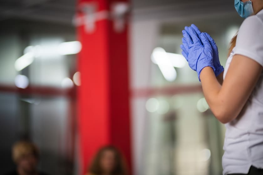
Our highly qualified and experienced trainers, above all, teach overcoming fear and take control of emotions that interfere with providing qualified assistance. The distinguishing characteristic of our trainings is a practical play component, which allows working out the necessary algorithms of actions and skills in a few hours until they become automatic. The friendly and supportive atmosphere allows our interns to relax and focus on the training materials.

Upon completion of each of the trainings, each participant who has mastered the necessary knowledge and skills, as well as successfully passed the tests, will receive a certificate. The type of the certificate depends on the training an attendee has chosen.
Continuous professional education of doctors
We constantly organize professional development activities: trainings, master classes, thematic professional schools, online courses for Dobrobut doctors. Our doctors may participate in internal and international traineeships, conferences and other professional events.
In addition to the development in the field of the clinical practice, we take care of upgrading the soft skills of our specialists: communications with patients, service, managing functions, mentoring.
Dobrobut Academy Medical School Research Center

Leading medical schools around the world have traditionally been research centers. We want this tradition to take root in Ukraine as well.
Research activities are a key focus area for Dobrobut Academy Medical School. Research is an integral part of medical training. Dobrobut Academy Medical School is the first and only medical school in Ukraine that combines research, teaching and practice to give its medical interns (now) and medical students (planned in 2023) access to the best medical education that prepares them for actual clinical practice. Our teaching staff engage in world-level scientific research to provide our medical interns and students with the most modern medical education.
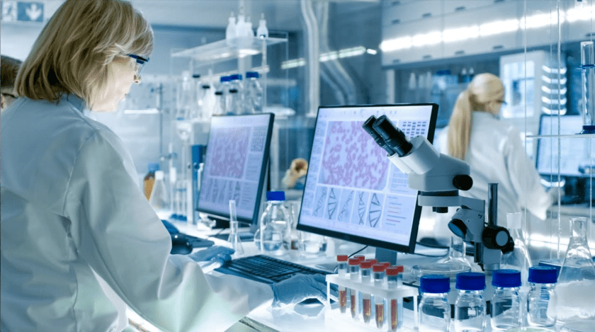
In this context, we are actively developing the Research Center of the Dobrobut Academy Medical School - a key element in the development of full-fledged university research. The Research Center includes laboratories of bioengineering, medical engineering and pain research, a behavioral tests module, molecular biology module, as well as core facilities, a vivarium, seminar rooms, and a recreation area.
Dobrobut Academy is open for cooperation with the media!
- Iryna Navolneva, head of the Dobrobut press center.
- Contacts: [email protected]
- Go to the Dobrobut Academy website to learn more about all the projects.

